.jpg)
Minimally Invasive
Patient-Matched
Solutions
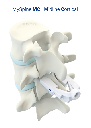
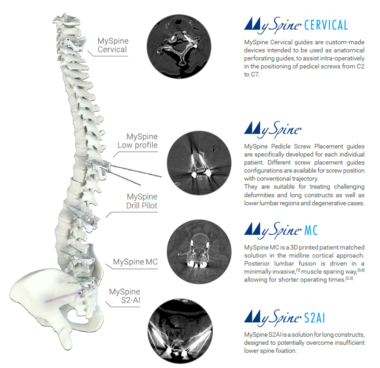
MySpine MC is a 3D printed patient matched solution in the midline cortical approach. Posterior lumbar fusion is driven in a minimally invasive [1,2,3], muscle sparing way, allowing for shorter operating times [11] and a substantial reduction of both radiation exposure [11] and costs [12].
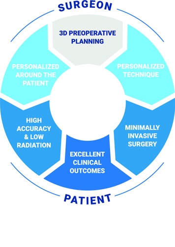
The goal of MySpine MC is to combine an excellent fusion rate with greater predictability of the clinical outcomes.
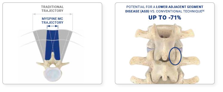
Minimally disruptive, medialized access with paramedial muscle retraction promotes[1]:
Enhanced muscle preservation[2]
Reduced blood loss[3]
Faster patient recovery[3]
Supradjacent facet preservation[1]

Entry points are located at the pars interarticularis with favourable cortical bone[4].

Improved bone purchase vs. conventional technique:
Ready to use 3D printed technology in your hands
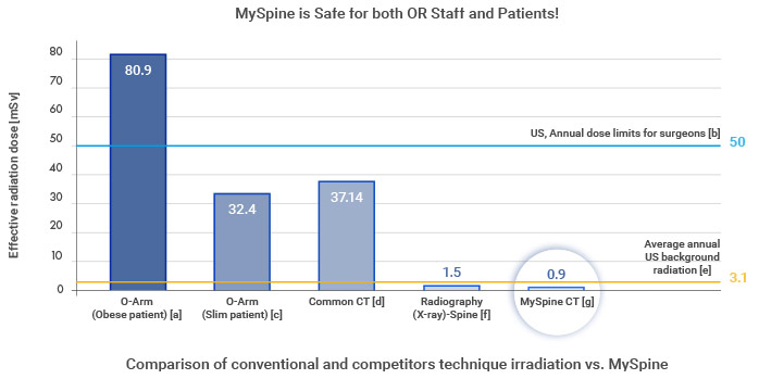
Patients are exposed to a low dose pre-op CT scan, resulting in radiation exposure lower than a single full spine x-ray
Pre-operative planning potentially nullifies the need of intra-operative checks, with dramatic reduction of irradiation[11,13]
Cumulative dose is potentially reduced vs. navigation assisted technique
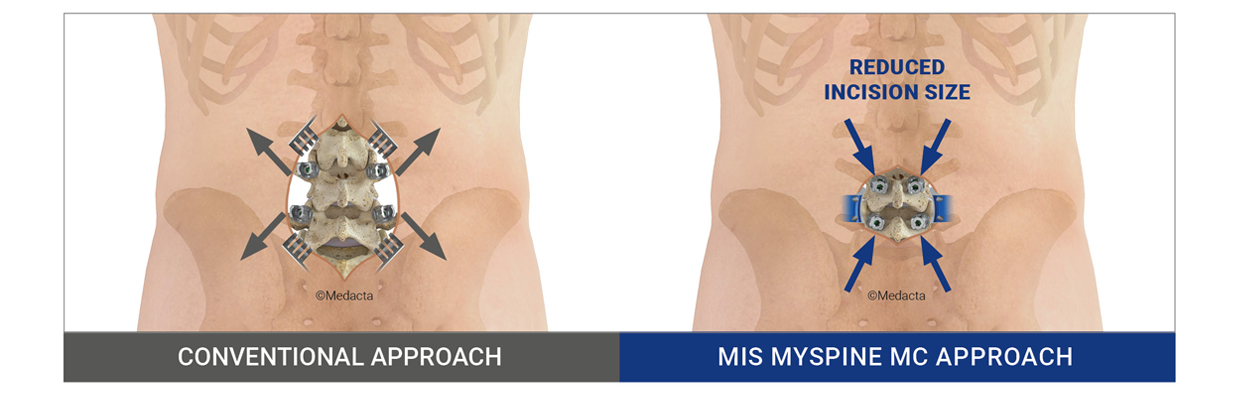
MySpine MC is truly a Minimally Invasive surgery [15,16]
Thanks to its muscle sparing technique, muscles are gently manipulated and a small skin incision of 4-5cm is performed.For this reason, MySpine MC represents an optimal system with its minimally disruptive atraumatic surgery, which is fundamental to deliver a fast recovery: MySpine MC will improve the patients quality of life and hasten their recovery after a spinal fusion surgery.
Why a MySpine MC Minimally Invasive surgery?
A Minimally Invasive surgery causes less surgical trauma than other techniques because back muscles are preserved, leading to a faster recovery. [15,16,17] Consequently, MIS MySpine MC approach can potentially provide you with the following benefits:

In comparison with “conventional” open surgical techniques, the MySpine MC approach can reduce the postoperative pain thanks to a less invasive technique. [16,17]
“Thanks to this surgery I have my life back.” A patient of MD P. Verstraete, Belgium

The MySpine MC technique can decrease the muscular atrophy leading to a potentially shorter rehabilitation, subject to doctor’s approval, based on the postoperative conditions. [16,17]
“My patients can walk autonomously the day after the surgery.” MD I. LaMotta, USA

The MySpine MC technique usually significantly reduces the duration of hospital stay. The surgeon may still recommend a longer stay, depending on the postoperative condition. [16,17]
“The patients treated with MySpine MC can leave the hospital on the 2nd postoperative day.” MD N. Marengo, Italy

With MySpine MC, the skin incision is often shorter than with “conventional” open surgery and therefore scar tissue is reduced. [16,17]

The MySpine MC 3D Printed Patient-Specific Solution can potentially provide better biomechanical performance, allowing for an improved long-term outcome. [15,16,17]
“At 6-month follow-up, our patients show important clinical improvements, without new neurologic deficits or radiologic pathologic findings.” MD K. Matsukawa, Japan

Preservation of muscles and vessels potentially reduces blood loss during the surgery. [16,17]

The MySpine MC technique reduces the incidence of complications when compared to free-hand techniques because of the highly accurate implant positioning.[19]
The MySpine MC Case management
MIS MySpine MC is a surgical instrument designed to accurately fit your vertebrae.

Medacta developed a specific Low Dose CT protocol to ensure a safe image acquisition. In fact the patient will receive a very similar amount of irradiation to only a single spine X-ray!

Using images of the patient spine, Medacta will create a plastic 3D model for each of the vertebra to be treated, in order to allow the Surgeon to select the best implant position and size.
.jpg)
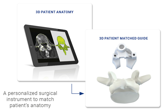
Using the model of the patient vertebrae and a dedicated planning software, the surgeon can tailor personalized surgical guides around each patient unique anatomy.
Prior to the surgery, surgeons will receive the MIS MySpine MC instruments and the plastic replica of the patient vertebrae. The bone model and the screw placement guides can be utilized to analyzed and accurately prepare for each spinal operation.

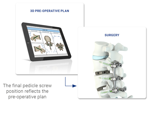
Surgeons will benefit of the MIS MySpine MC guides that will help them positioning the pedicle screws very accurately according to pre-operative plan.
Thanks to this accurate tool the surgeon can optimize screws parameters, entry points and trajectories[14], avoiding potential intraoperative complications for the patient, such as pedicle fractures and neurovascular injuries[14,16].
MySpine MC entry points and trajectories are customized through pre-op trajectory management to enable the use of longer screws and larger diameters vs. free hand CBT, and are comparable to the conventional technique.
Following the pre-op trajectory a 3D patient matched guide is designed to match the patient’s anatomy. This navigated tool provides accurate intra-operative guidance for safe screw positioning[14] potentially reducing the need of fluoroscopy[15].
A comprehensive range of patient specific, pedicle screw placement guides allows for a personalized treatment depening on the patient pathology and the surgical approach. The system supports the surgeon pre and intra operatively for post op patient benefit.
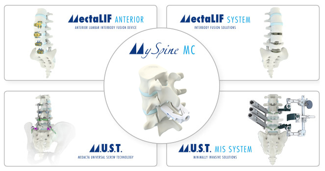
MySpine MC, together with the MUST Screw System, the MectaLIF Family of Interbody Fusion Devices and our Suite of Specialized Surgical Instruments, create a harmonized and complete system

MyKnee is a patient-specific cutting block, allowing the surgeon to realize his pre-operative 3D planning, based on CT or MRI images of the patient’s knee.
This innovative concept combines different features giving potential benefits to both the surgeon and to the patient.

MyHip is a system providing 3D preoperative planning and patient-specific guides, developed following the success of Medacta Patient Matched Technology

MyShoulder is a patient-specific instrumentation, allowing the surgeon to realize his pre-operative 3D planning, based on CT images of the patient’s shoulder. The pre-operative 3D planning allows to manufacture an humeral resection guide and a glenoid positioning guide.
This innovative concept combines different features giving potential benefits to both the surgeon and to the patient.
An advanced network of digital solutions designed to improve patient outcomes and healthcare efficiency
[1] Matsukawa K. et al., Incidence and Risk Factors of Adjacent Cranial Facet Joint Violation Following Pedicle Screw Insertion Using Cortical Bone Trajectory Technique, Spine, 2016
[2] Sakaura H. et al., Posterior lumbar interbody fusion with cortical bone trajectory screw fixation versus posterior lumbar interbody fusion using traditional pedicle screw fixation for degenerative lumbar spondylolisthesis: a comparative study, JNS, 2016
[3] Khanna N. et al,. Spine (Phila Pa 1976). 2016 Apr;41 Suppl 8:S90-6. doi: 10.1097/BRS.0000000000001475. Medialized, Muscle-Splitting Approach for Posterior Lumbar Interbody Fusion: Technique and Multicenter Perioperative Results
[4] Gautschi O. et al., Maximal access surgery for posterior lumbar inter body fusion (PLIF) with divergent, cortical bone trajectory (CBT) pedicle-screws: a good option for minimize spine access and maximize the field for nerve decompression, Journal of neurosurgical sciences, 2015
[5] Matsukawa -2nd MORE Japan MySpine cortical Bone Trajectory 2017.
[6] Matsukawa - Biomechanics of CBT (internal file)
[7] Regev G etal., Nerve injury to the posterior rami medial branch during the insertion of pedicle screws: comparison of mini-open versus percutaneous pedicle screw insertion techniques. Spine. 20093411239-42
[8] Lamartina C. et al., Pedicle screw placement accuracy in thoracic and lumbar spinal surgery with a patient-matched targeting guide: a cadaveric study, European Spine Journal, 2015
[9] Santoni B.G. et al., Cortical bone trajectory for lumbar pedicle screws, The Spine Journal, 2009
[10] Mori K. et al., Short-Term Clinical Result of Cortical Bone Trajectory Technique for the Treatment of Degenerative Lumbar Spondylolisthesis with More than 1-Year Follow-Up, Asian Spine Journal, 2016
[11] Farshad M. et al., Accuracy of patient-specific template-guided vs. free-hand fluoroscopically controlled pedicle screw placement in the thoracic and lumbar spine: a randomized cadaveric study, European Spine Journal, 2017
[12] Chin K.R., Clinical Outcomes With Midline Cortical Bone Trajectory Pedicle Screws Versus Traditional Pedicle Screws in Moving Lumbar Fusions From Hospitals to Outpatient Surgery Centers, Clinical Spine Surgery, 2017
[13] Kaito T., Cortical pedicle screw placement in lumbar spinal surgery with a patient-matched targeting guide: A cadaveric study, Journal of Ortopaedic Science, 2018
[14] Matsukawa K. et al., Accuracy of cortical bone trajectory screw placement using patient-specific template guide system, Neurosurgical Review, July 2019
[15] Matsukawa K. et al., Cortical pedicle screw trajectory technique using 3D printed patient-specific-guide, M.O.R.E. Journal, September 2018.
[16] Marengo N. et al., Cortical Bone Trajectory Screw Placement Accuracy with a Patient-Matched 3-Dimensional Printed Guide in Lumbar Spinal Surgery: A Clinical Study, WORLD NEUROSURGERY, June 2019
[17] Marengo N. et al., Cortical Bone Trajectory Screws in Posterior Lumbar Interbody Fusion: Minimally Invasive Surgery for Maximal Muscle Sparing—A Prospective Comparative Study with the Traditional Open Technique, Clinical Study, February 2018
[18] Kim J. et al., Three-Dimensional Patient-Specific Guides for Intraoperative Navigation for Cortical Screw Trajectory Pedicle Fixation, World Neurosurgery Volume 122 (674-679), February 2019
[19] Petrone S. et al., Cortical bone trajectory technique’s outcomes and procedures for posterior lumbar fusion: A retrospective study, Journal of Clinical Neuroscience, April 2020
[a] Lange et.al. Estimating the effective radiation dose imparted to patients by intraoperative cone-beam computed tomography in toracolumbar spinal surgery, Spine 2013
[b] US Nuclear Regulatory Commission’s (USNRC)
[c] Lange et.al. Estimating the effective radiation dose imparted to patients by intraoperative cone-beam computed tomography in toracolumbar spinal surgery, Spine 2013
[d] Biswas et.al. Radiation Exposure from Musculoskeletal Computerized Tomographic Scans, JBJS Am. 2009
[e] Health Physics Society Specialists in Radiation Safety, Lawrence Berkeley National Laboratory; Fact Sheet 2010
[f] Radiation Dose in X-Ray and CT Exams; 2013 Radiological Society of North America, Inc
[g] MySpine, Charité University Hospital, Berlin, Germany